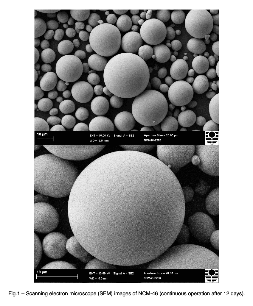
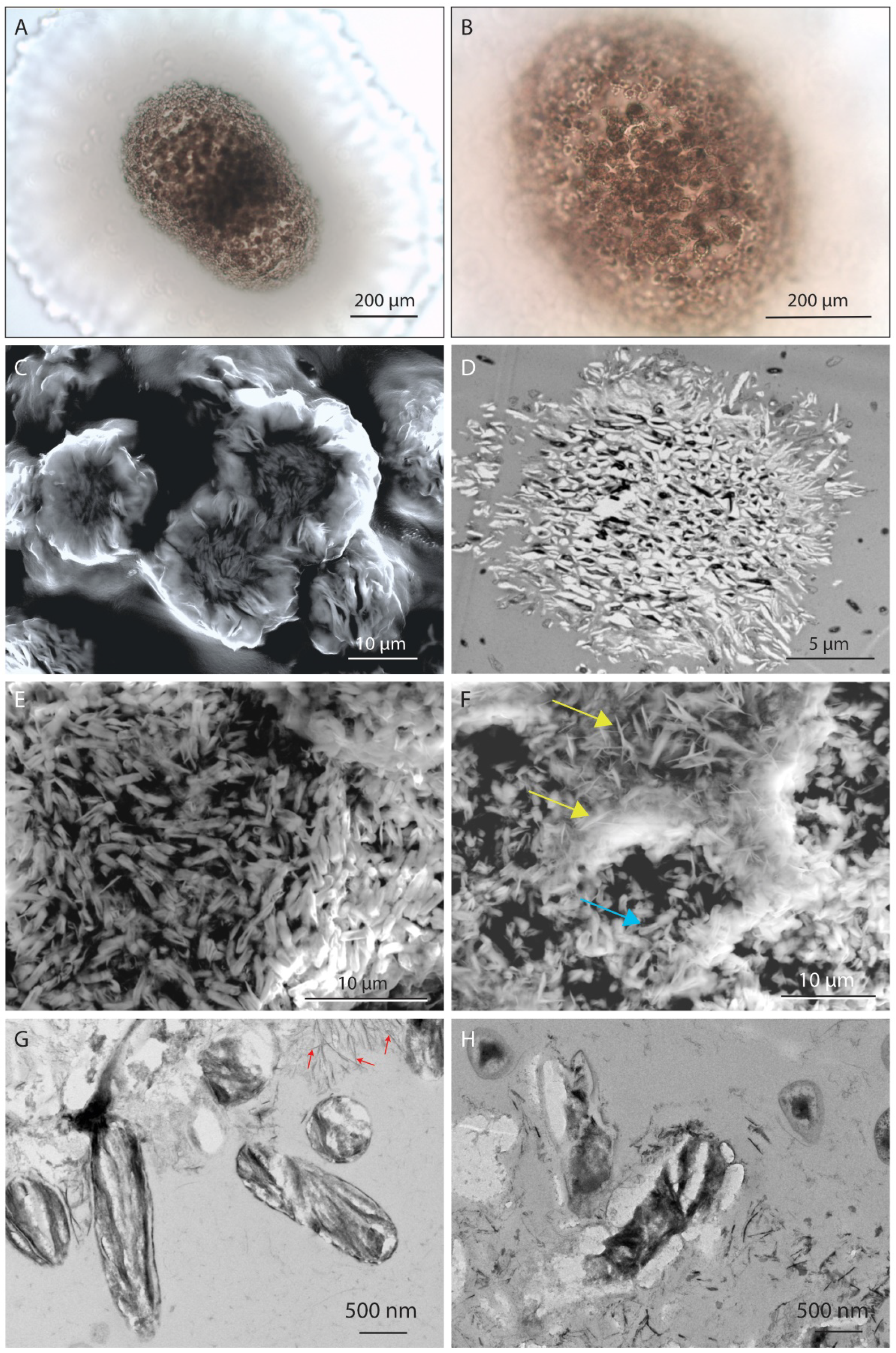
SEM (Scanning Electron Microscope) microphotographs of manganese
Download scientific diagram | SEM (Scanning Electron Microscope) microphotographs of manganese micronodules from the depth of 300 to 305 cm, size fraction 100-250 μm: а - micronodule with the frustules of Ethmodiscus, б - micronodule without admixture of valves of Ethmodiscus. from publication: Anomalies of rare elements in manganese micronodules from ethmodiscus oozes in the Brazil basin of the Atlantic Ocean | The composition of manganese micronodules from miopelagic clays and Ethmodiscus oozes of the central part of the Brazil Basin (station 1537, R/V Akademik Sergei Vavilov) is considered. Micronodules were recovered from >50 μm fraction of sediments from the depth intervals of | Manganese, Brazil and Atlantic Ocean | ResearchGate, the professional network for scientists.

Photomicrographs - an overview

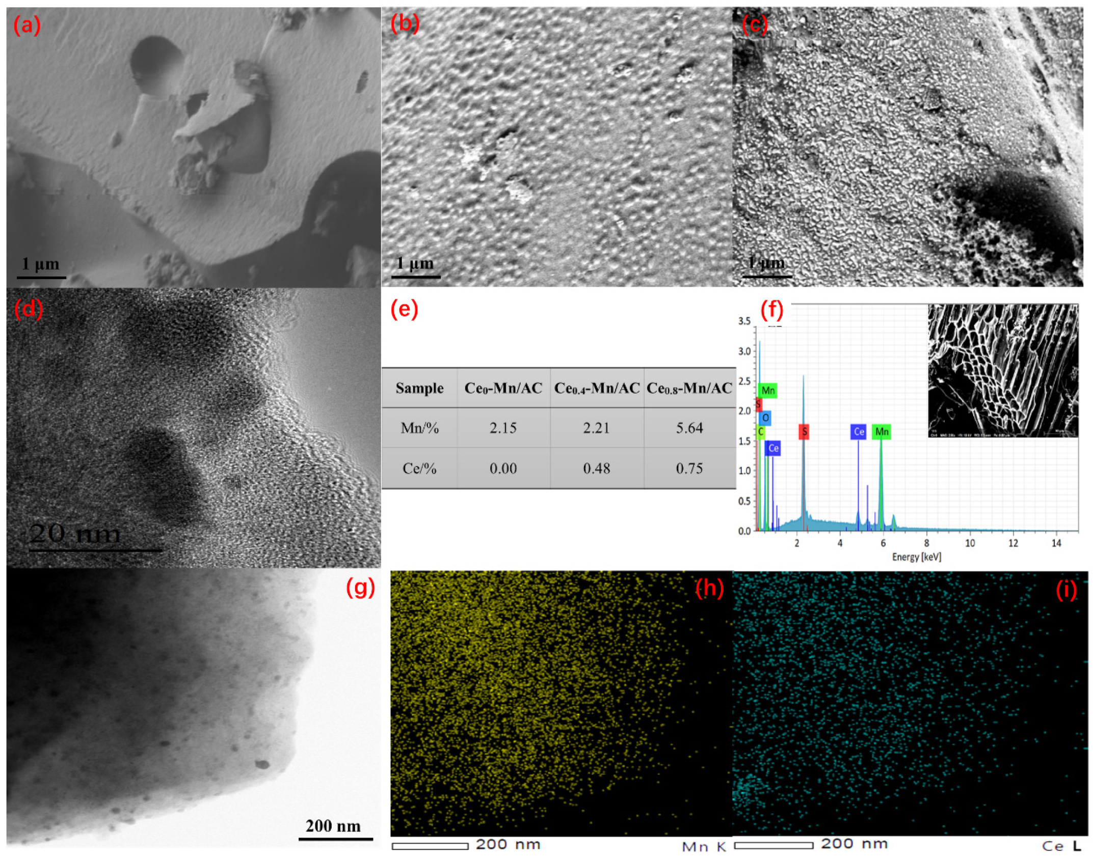
Scanning electron microscope (SEM) images with EDS elemental maps
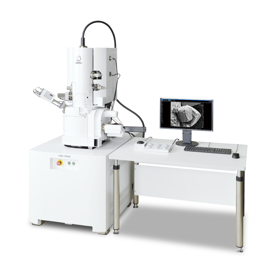
JSM-IT800 Schottky Field Emission Scanning Electron Microscope

Figure S2. Scanning electron microscopy (SEM) images of manganese

Experimental methods in chemical engineering: Scanning electron
Investor News - Manganese Sulphate MnSO4 - Pilbara Metals Group

Scanning electron microscope micrographs of the black deposit of

Scanning Electron Microscope, Apreo 2 SEM
IJERPH, Free Full-Text

Scanning Electron Microscopy

Scanning electron microscope (SEM) images, secondary electron mode
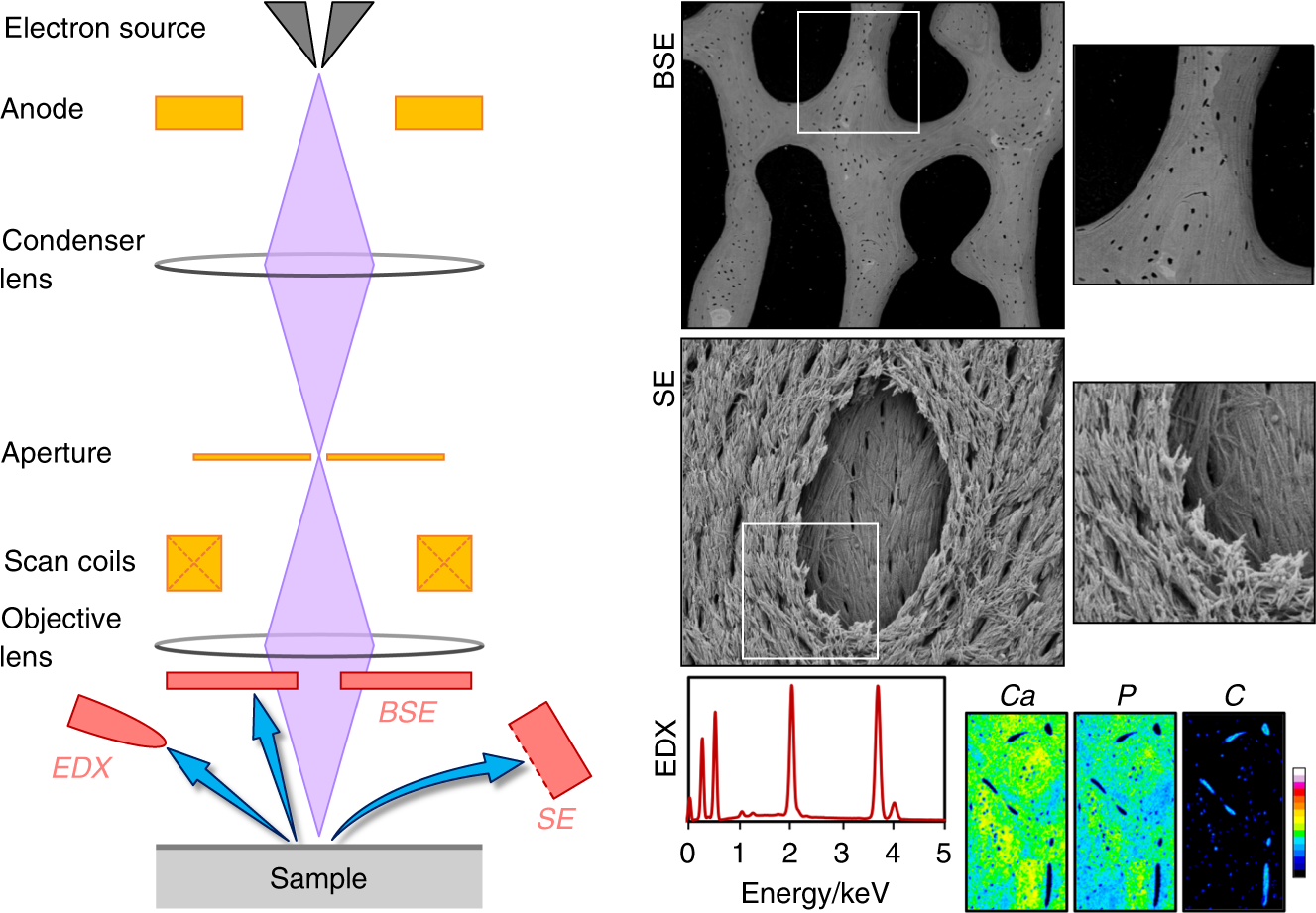
50 years of scanning electron microscopy of bone—a comprehensive
Minerals, Free Full-Text

Scanning electron microscopy (SEM) images of carbon nanotubes

Synthesis and characterization of ZnS:Mn/ZnS core/shell